Vision dysfunction following a brain injury
Having recently had my first eye test since primary school and my first pair of glasses, I started thinking about the common visual problems experienced by a large proportion of the population and the more complex visual problems often experienced following a brain injury.
Many people will be familiar with the common types of vision and vision problems with almost 60% of people in the UK being affected by visual impairments and requiring glasses or contact lenses.
The common types of vision in technical terms are:
Emmetropia: This means you have ideal distance vision or normal vision
Near-sighted: Where objects up close look visibly clear but objects in the distance are blurry you are must likely near-sighted also referred to as short-sighted and would benefit from wearing glasses or contact lenses
Farsighted: If objects up close are blurry, but you can see objects at a distance clearly, you are farsighted or longsighted. This problem can also be corrected with glasses or contact lenses.
Astigmatism: If you experience blurred or distorted vision at any distance in one or both eyes, you most likely have astigmatism. This condition affects almost half of the population and can affect your ability to focus on objects resulting in headaches and fatigue. This condition can be corrected with glasses or contact lenses or laser surgery.
Presbyopia: This is the gradual loss of your eye’s ability to focus on nearby objects and usually occurs with age. Wearing glasses is the easiest way to correct this problem.
Many of us are or will experience one of the above problems in our life time or know someone is. There are other less common visual dysfunctions affecting a smaller percentage of the population; often those who have suffered a head or brain injury. I will explore some of these below.
Visual dysfunction following a brain injury
We get 80-90% of our sensory input from vision. Our brains needs vision to decode information that it receives from the eyes so to make sense of our environment. The brain requires different aspects of vision to process and recognise different shapes, colours, faces and words and to process the movement of objects or location/position of an on object.
The ability to process these aspects of vision are generally governed by different parts of the brain therefore an injury to the brain can affect any aspect of vision.
Below are some of the common vision problems which a person who has suffered a brain injury may experience.
Visual field loss
Following a brain injury parts of the visual field may be affected or lost. This can result in missing patches of vision often appearing as black or fuzzy patches. Vision may also be lost around the edges or centre of the eye. The term hemianopia refers to loss of half of your visual field while quadrantanopia refers to loss of a quarter of the visual field. Someone who is suffering from visual field loss can undergo vision compensatory training to help you use their remaining vision more efficiently or vision restoration therapy to improve visual information processing.
Diplopia
Diplopia also known as double vision causes a person to see two images of one single object. Where this occurs in one eye it is known as monocular diplopia and if it occurs in both eyes it is known as binocular diplopia. Diplopia can sometimes correct itself following an injury and if so usually within approximately three months. There are also a number of potential treatments for diplopia including eye exercises, wearing an eye patch or opaque contact lenses over the affected eye, Botox injections into the eye muscle so as to help them relax or surgery to correct the positioning of the eye muscles.
Photophobia
This is an extreme sensitivity to light and is a commonly reported visual disturbance amongst brain injury survivors. The sensitivity can be such that it causes discomfort or even pain. This condition can make it very difficult to go to certain places where the light is too bright. Research shows that in approximately half of cases of photophobia caused by brain injury, the condition will improve over time. Treatment for photophobia caused by a brain injury includes wearing tinted glasses and getting botox injections.
Stereopsis
Also known as depth perception, this condition results in the eyes being unable to measure the distance between objects. A deficiency in stereopsis can result in a lack of spatial awareness and difficulty with all sorts of everyday activities such as driving or climbing the stairs. Where this condition occurs following a brain injury it will often be due to cortical damage. Treatment depends on the cause and management includes visual exercises and therapy and in some cases specialised glasses.
Nystagmus
This is a form of visual impairment, characterised by involuntary rhythmic shaking of the eyes, from side to side, up and down or round and round. This affects the ability to focus, judge speed and depth and recognise faces. Nystagmus is caused by the abnormal functioning of the part of the brain or inner ear which regulates eye movement and positioning and can cause nausea and vertigo. Where Nystagmus occurs after a brain injury it is known as acquired nystagmus. In some cases acquired nystagmus can come and go over time and sometimes symptoms can reduce or disappear on their own. Certain drug treatments can be used to try and reduce the effects of acquired nystagmus as well as Botox (botulinum) injections.
Diagnosing and treating visual dysfunction following a brain injury
Visual problems following a brain injury can manifest themselves as:
- Postural changes, for example sitting at an angle
- Slow, clumsy movements or coordination difficulties
- Headaches, dizziness or fatigue caused by eye strain
- Vertigo or nausea
- Anxiety and avoiding social interaction
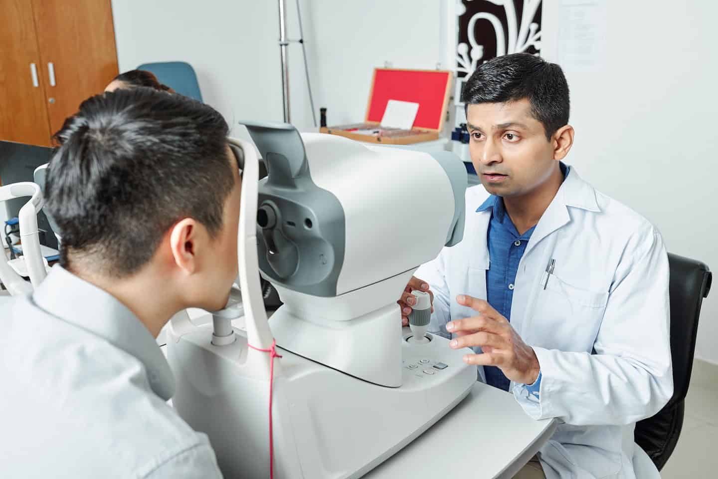
Once difficulties have been detected, they should be assessed by a clinician so as to identify whether they relate to an injury to the brain or eye(s). This can be a complex process due to the complexity of the visual pathway between the eyes and the brain therfore it is important to seek treatment from a clinician qualified to diagnos/treat your visual problems.
Neuro-ophthalmologist: This is a medical doctor specifically trained in the medical and surgical care of eyes and specialising in the brain’s role in the visual system. Ideally someone who is experiencing visual dysfunction following a brain injury would be assessed by a neuro-ophthalmologist where their problems are complex.
Orthoptist: This is a medical professional who primarily assesses and treats eye muscle disorders
Optmetrist: An optometrist is a specialist who tests eyes and prescribes visual aids such as glasses and contact lenses
Optician: Often confused for an optemetrist, an optician fits glasses and contact lenses
It may sometimes also be appropriate to use neuroimaging tools such as CT and MRI scans to examine the brain to help identify the damage causing the visual defects. This is something you should query with your doctor if your visual problems remain undiagnosed following examinations with the above experts.
Help and advice
To find out more about visual problems following a brain injury, you can visit Headway, the brain injury association dedicated to improving life after brain injury where you will find further information, useful links and advice.
If you or someone you know are suffering from nystagmus, visit the Nystagmus Network for support and information.
To access free online therapy for those experiencing visual search problems following a brain injury, visit Eye-Search Therapy.
